

ALBUM
Automated Large-scale Brain Ultrastructure Mapping
Abstract
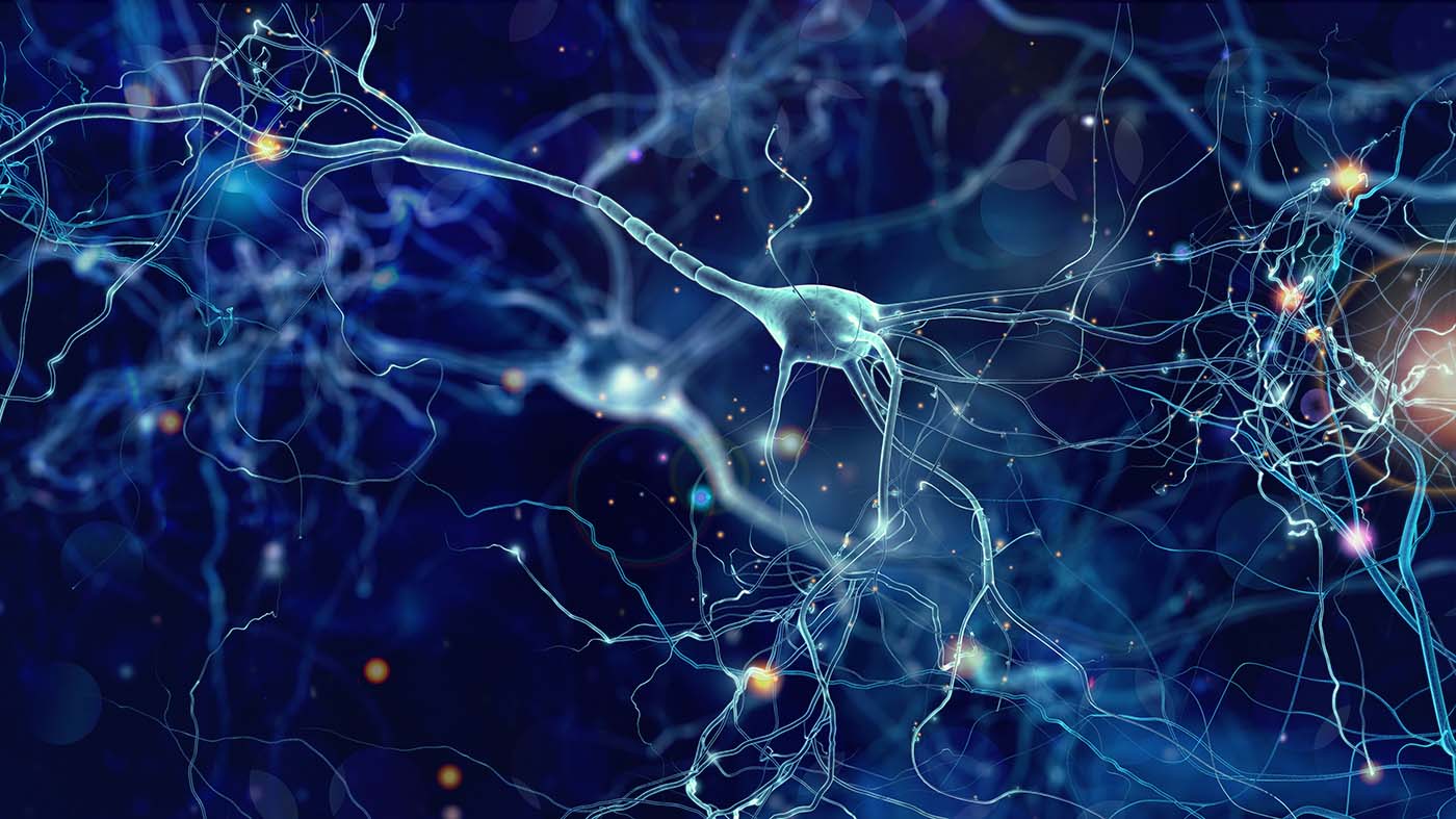
Mapping connections between neurons requires detailed 3D images of brain tissue and accurate identification of individual neurons. Conventional electron microscopy creates these images from thousands of ultra-thin sections, but missing or damaged sections can distort the results and cause segmentation errors. This project investigates a more robust approach: repeatedly imaging thicker tissue sections while removing very thin layers between scans. We develop computational methods to reconstruct detailed 3D images from these measurements and identify imaging strategies that maximize information without increasing experimental costs. We also improve automated 3D neuron segmentation by combining advanced deep learning with knowledge of biological structures. Early results show that this refinement improves segmentation accuracy. The expected outcome is a more reliable and efficient pipeline for reconstructing brain tissue and mapping neuronal connections.
People
Collaborators

Suman pursued his doctoral studies in the Visual Artificial Intelligence Laboratory at Oxford Brookes University, United Kingdom, focusing on spatiotemporal human action localization using deep learning techniques. Suman received a Ph.D. in Computer Science and Mathematics from Oxford Brookes University in 2017. He served as a postdoctoral fellow at Oxford Brookes University between December 2017 and July 2018. Then he moved to CVL (Computer Vision Lab) at ETH Zurich, holding a postdoctoral researcher position until June 2023. Suman's research has centered around unsupervised domain adaptation (UDA) for visual scene understanding (semantic and panoptic segmentation), human behavior understanding, and vision-based biometrics (face anti-spoofing). He also worked on semi-supervised learning for semantic segmentation by leveraging self-supervised depth estimation. His internship at Disney Research Zurich involved designing deep generative models for unsupervised facial expression learning. Additionally, Suman tackled research problems in multi-task learning (MTL) by addressing two common challenges in developing multi-task models, incremental learning and task interference.



Aleix Boquet i Pujadas joined the SDSC in October 2025 as a Senior Data Scientist in the Paul Scherrer Institute (PSI) team, based in Villigen.
He received his BSc in Math and Physics by the Universtat Autònoma de Barcelona (2016), and his PhD in EE&CS from Université Sorbonne (2019) working on Biomedical Image Analysis. He has been a R&D Associate at Institut Pasteur (2020-2021) and postdoctoral fellow at the Biomedical Imaging Group at EPFL (2021-2025). Aleix comes with strong expertise in optimization and inverse problem modeling with a focus on biomedical imaging applications.

Benjamín Béjar received a PhD in Electrical Engineering from Universidad Politécnica de Madrid in 2012. He served as a postdoctoral fellow at École Polytechnique Fédérale de Lausanne until 2017, and then he moved to Johns Hopkins University where he held a Research Faculty position until Dec. 2019. His research interests lie at the intersection of signal processing and machine learning methods, and he has worked on topics such as sparse signal recovery, time-series analysis, and computer vision methods with special emphasis on biomedical applications. Since 2021, Benjamin leads the SDSC office at the Paul Scherrer Institute in Villigen. Since 2025, he serves as Domain Lead for Large-Scale Scientific Infrastructures.
PI | Partners:
PSI, Structural Neurobiology Group, Laboratory of Multiscale Bioimaging (LMB)
- Dr. Adrian Andreas Wanner (Group Leader)
description
Motivation
Understanding the detailed wiring of the brain requires imaging and analyzing neural tissue at nanometer-scale resolution over very large volumes. Volume electron microscopy (vEM) has become a key technology for this purpose, as it can reveal cellular and subcellular structures with the resolution needed for connectomics and the study of healthy and diseased brain tissue. However, current vEM workflows remain difficult to scale. Conventional serial-section methods rely on cutting thousands of ultrathin sections, typically 30–40 nm thick, and collecting them reliably. This process is slow, technically demanding, and highly sensitive to artifacts such as section loss, cracks, folds, distortions, and insufficient axial resolution. These artifacts directly affect the quality of reconstructed 3D volumes and are among the main reasons why automated neuron segmentation still produces errors such as over-merging and over-segmentation.
The motivation of this project is to develop a more scalable computational imaging and analysis pipeline for large-scale brain tissue reconstruction. By using semi-thin sections combined with iterative broad ion-beam milling and cumulative transmission imaging, we aim to reduce the dependence on fragile ultrathin sectioning while still recovering high-resolution 3D information. This creates an important computational challenge: the acquired images are cumulative projections through the remaining sample, so recovering the underlying 3D volume requires solving an ill-conditioned inverse problem. At the same time, the reconstructed volumes must be suitable for downstream automated neuron segmentation, which is essential for biological interpretation.
Therefore, this project is motivated by the need to jointly address reconstruction and segmentation rather than treating them as independent problems. Improved reconstruction can provide cleaner 3D volumes, while robust deep-learning-based segmentation can transform these volumes into biologically meaningful neural structures. By combining inverse-problem-based reconstruction, optimized acquisition design, and biologically informed deep-learning segmentation, the project aims to enable more accurate, efficient, and scalable analysis of large vEM datasets for connectomics and neuroscience.
Proposed Approach / Solution
The proposed solution is a complete computational pipeline that addresses both 3D reconstruction and 3D neuron segmentation for cumulative volume electron microscopy. The first part of the approach focuses on reconstructing high-resolution 3D volumes from cumulative transmission measurements acquired during iterative layer removal. In this imaging setup, semi-thin sections are gradually milled using a broad ion beam, and after each milling step, a projection image is acquired through the remaining sample using multibeam scanning transmission electron microscopy. Since each image contains cumulative information from the remaining layers, recovering the underlying 3D structure is formulated as an inverse problem. We model the image formation process using a cumulative-sum forward operator and develop optimization-based reconstruction strategies to recover the individual layers from the projection sequence. To address the ill-conditioning of the inverse problem and detector noise, we use linearization and regularized optimization methods. In addition, we investigate acquisition design by identifying maximally informative layer-removal patterns, so that better reconstructions can be obtained under the same experimental budget.
The second part of the approach focuses on automated 3D neuron segmentation from the reconstructed EM volumes. We establish two strong deep-learning baselines: CAD, which uses cross-dimension affinity distillation to transfer 3D structural knowledge to an efficient 2D student network, and AGQ, which uses affinity-guided queries to directly predict neuron segmentations more efficiently than traditional watershed-based pipelines. Building on these baselines, we propose a biologically informed refinement strategy that improves AGQ segmentation outputs using a biological graph representation. This graph-based refinement incorporates structural consistency and biological plausibility into the final segmentation, helping to reduce over-merging and over-segmentation errors.
Overall, the proposed approach combines inverse-problem-based 3D reconstruction, information-theoretic acquisition optimization, and deep-learning-based neuron segmentation. This integrated solution is designed to produce high-quality 3D EM volumes and more accurate neuron segmentations for scalable connectomics analysis.
Impact
The expected impact of this project is to make large-scale volume electron microscopy more accurate, efficient, and scalable for brain connectomics. By reducing the dependence on fragile ultrathin serial sectioning, the proposed imaging and reconstruction pipeline can lower artifact-related failures and improve the quality of 3D EM volumes. At the same time, improved deep-learning-based neuron segmentation can reduce over-merging and over-segmentation errors, enabling more reliable analysis of neural circuits. Together, these advances can support petabyte-scale reconstruction and segmentation of brain tissue, helping researchers better study ultrastructural changes in healthy and diseased brains.
Presentation
Gallery
Annexe
Additional resources
Bibliography
- CAD: Liu, X., Cai, M., Chen, Y., Zhang, Y., Shi, T., Zhang, R., ... & Xiong, Z. (2024, June). Cross-dimension affinity distillation for 3d em neuron segmentation. In 2024 IEEE/CVF Conference on Computer Vision and Pattern Recognition (CVPR) (pp. 11104-11113). IEEE Computer Society.
- AGQ: Chen, H., Tang, C., Li, X., & Hu, X. (2025). Efficient Neuron Segmentation in Electron Microscopy by Affinity-Guided Queries. In The Thirteenth International Conference on Learning Representations.
- Biologically-Constrained Graphs: Matejek, B., Haehn, D., Zhu, H., Wei, D., Parag, T., & Pfister, H. (2019). Biologically-constrained graphs for global connectomics reconstruction. In Proceedings of the IEEE/CVF conference on computer vision and pattern recognition (pp. 2089-2098).
Publications
Related Pages
More projects

AICADES
IICU National Data Stream
SMARTT

TGSF-DA
More projects
News
Latest news

Quand l’IA soutient l’économie du canton de Vaud, la santé et l’environnement
Quand l’IA soutient l’économie du canton de Vaud, la santé et l’environnement

Elevating International Geneva's Impact with AI
Elevating International Geneva's Impact with AI
12 Zurich SMEs join next AI Innovation Program
12 Zurich SMEs join next AI Innovation Program
Contact us
Would you like to explore how we can work together?
Our team of data scientists and professionals looks forward to engaging in impactful, results-driven collaborations with you.